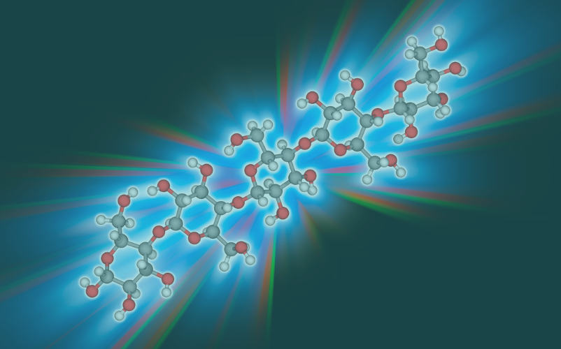
The biochemical structure of LPS and how it acts in nature vs. the lab
The cell envelope functions as the outermost barrier, shielding bacteria from their external surroundings. The makeup and architecture of a bacteria's outer layer are crucial for how it interacts with things like viruses, other bacteria, the immune system, and even chemical attacks. Lipopolysaccharide (LPS), a sugar-lipid compound that coats the exterior of many Gram-negative bacteria, is a prime target for scientific investigation. [1]
Structure of Bacterial Cell Wall
The defining characteristic of Gram-negative bacteria is their cell envelope, which features two membranes: an inner membrane (IM) enclosing the cytoplasmic contents and an outer membrane (OM) separating the cell from its environment. Most Gram-negative bacteria possess an outer membrane (OM) that does not exhibit a phospholipid bilayer structure. Instead, the bilayer exhibits high asymmetry, with phospholipids comprising the inner leaflet and lipopolysaccharide (LPS) molecules composing the outer leaflet.[2]
Biochemical structure of LPS
LPS is a large glycolipid, consisting of three distinct parts: the fat-like lipid A, a complex sugar chain called the core oligosaccharide, and the O antigen, which vary between different bacteria. Lipid A, the molecule's hydrophobic part, is a glucosamine disaccharide with acylated β-1′-6 linkages that makes up the outer layer of the outer membrane (OM). The core oligosaccharide, attached to lipid A's glucosamines, is unique and doesn't repeat. The O antigen, a complex polysaccharide, is bound to the core oligosaccharide.[1, 3]
How LPS acts?
The LPS's pathogenicity mainly comes from Lipid A, which was previously called catechin. Even in small amounts, Lipid A can trigger fever and metabolic changes, indicating its strong pathogenicity. Individuals infected with lysed Gram-negative bacteria may experience a rapid increase in body temperature (fever). The cellular network's breakdown of Gram-negative bacteria liberates Lipid A when cell walls rupture, enabling its binding to effector cells, including macrophages. Fatal doses may cause septic shock, low blood pressure, and fever. The action of Lipid A on macrophages results in the production of pyrogens, leading to the activation of mediators like prostaglandins.[4]
The impact of LPS on various laboratory tests.
A wide range of lab tests are used to detect endotoxin contamination in medical devices and drugs, providing crucial information about their safety. The confirmation of contamination is established by the reaction between the reagent and endotoxin.
RPT (Rabbit Pyrogen Test): The rabbit pyrogen test, the first method approved by the US Food and Drug Administration for LPS detection, relied on the simple principle of measuring an endotoxin's ability to induce fever in a rabbit, effectively gauging the toxin's potency.
LAL (Limulus Amoebocyte Lysate): The LAL assay, the gold standard for lipid A detection, can be inhibited by various mechanisms and is prone to variability. As a result, guidelines for manufacturing and testing assays for human products have been published by the United States Pharmacopeia and the Code of Federal Regulations. If bacterial endotoxin is present, the LAL assay detects it by setting off a series of enzymatic reactions that lead to clotting, which serves as a clear signal of contamination.[5]
MAT (Monocyte Activation Test): Because it uses human cells, the MAT more closely mimics the reactions and responses of a living organism. The MAT involves subjecting human monocytic cells to pyrogens in a controlled environment, triggering the activation of the cells. Proinflammatory cytokines are released as a result of this activation and can be detected through the ELISA technique.[6]
Effect of change in the molecular conformation of the LPS on endotoxin activity
Low endotoxin recovery (LER) raised concerns about the LAL assay's potential to miss LPS in specific situations. Inaccurate LPS detection jeopardizes experimental results and creates a safety hazard in industrial applications where LPS might go undetected.[7]
Classification of LPS based on structural changes
Whether LPS is classified as high (smooth) or low (rough) molecular weight depends on whether its structure includes an O-antigen. O-antigen absence is possible naturally or through genetic mutation. Although all rough mutants are devoid of O-antigen, the oligosaccharide core length differs based on the involved gene loci.
Each method detects LPS, but their activities vary. Studies indicate similar kinetic profiles for LAL and rFC, but MAT exhibited enhanced masked endotoxin recovery. Altering LPS affects masking regardless of the assay. Masking susceptibility is influenced by polysaccharide length: shorter chains (rough LPS) decrease masking due to their lower hydrophilic/hydrophobic ratios. Furthermore, the increased negative charge in rough mutants improves cation binding, which likely contributes to the stability of supramolecular assemblies, making them less prone to masking than smooth mutants.[8]
The reactions triggered by LPS in nature serve as the primary reference for creating accurate lab tests.
References:
1.Bertani, B. and N. Ruiz, Function and Biogenesis of Lipopolysaccharides. EcoSal Plus, 2018. 8(1).
2.Silhavy, T.J., D. Kahne, and S. Walker, The bacterial cell envelope. Cold Spring Harb Perspect Biol, 2010. 2(5): p. a000414.
3.Kalynych, S., R. Morona, and M. Cygler, Progress in understanding the assembly process of bacterial O-antigen. FEMS Microbiol Rev, 2014. 38(5): p. 1048-65.
4.Schultz, C.J.G.I.M.C.I., Lipopolysaccharide, structure and biological effects. 2018. 3(1): p. 1-2.
5.Loreen, R.S., M.M. Heather, and M. Harshini, Detection Methods for Lipopolysaccharides: Past and Present, in <i>Escherichia coli</i>, S. Amidou, Editor. 2017, IntechOpen: Rijeka. p. Ch. 8.
6.Perdomo-Morales, R., et al., Monocyte activation test (MAT) reliably detects pyrogens in parenteral formulations of human serum albumin. Altex, 2011. 28(3): p. 227-35.
7.Gorman, A. and A.P. Golovanov, Lipopolysaccharide Structure and the Phenomenon of Low Endotoxin Recovery. European Journal of Pharmaceutics and Biopharmaceutics, 2022. 180: p. 289-307.
8.Burgmaier, L., et al., The impact of LPS mutants on endotoxin masking in different detection systems. Biologicals, 2025. 89: p. 101808.


