Principles on which the Limulus Test for the Detection of Bacterial Endotoxins is Based
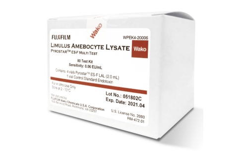
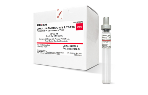
The LAL or Limulus test is used for the determination of bacterial endotoxins in a wide variety of samples in both research laboratories and industries. The principle of this test is based on the process of coagulation which occurs in the hemolymph of horseshoe crab (Limulus Polyphemus) in the presence of lipopolysaccharides. In this article, the biological process and the way it was used to develop the test is described.
Discover and quote online our Endotoxin-Specific LAL Reagents HERE
The coagulation of the hemolymph of the horseshoe crab occurs through a series of chain reactions similar to those observed in mammals. For these chain reactions to be activated, the serine protease pro-enzymes present in the hemolymph need to react successively to cause the gelation process. A significant characteristic feature of coagulation reactions occurring in the Limulus crab is that they are activated with very small amounts lipopolysaccharides (LPS); concentrations of bacterial endotoxins coming into contact with the hemolymph at the picogram level are already sufficient for the reactions to occur. These reactions have the function of immobilising pathogenic micro-organisms, which die due to antimicrobial substances secreted in parallel and released into the hemolymph.
The fact that many of the necessary precursors for clots to be formed in the hemolymph of the horseshoe crab are found in the hemolymph in the form of granules, known as amebocytes, was used by researchers to develop a method of analysis of bacterial endotoxins by extracting the hemolymph of the crab. This test is known as the Limulus Amebocyte Lysate (LAL) as the lysate of the granules is precisely performed so that they react to the presence of endotoxins in the test environment and gelation is produced. Gelation is the analytic signal used for both the qualitative and quantitative detection of LPS. To perform the test, an amount of hemolymph of each crab is extracted with a syringe and the animal is left in its environment.
FEATURED: The Detection of Endotoxins via the LAL Test, the Gel Clot Method
If we go more into the biological process that occurs in the hemolymph of the horseshoe crab in the presence of LPS, we can see a number of separate activation processes from pro-enzymes to proteolytic enzymes: - LPS activates the reaction of pro-enzyme factor C autocatalytically in its transformation to activated factor C; - The factor C, in turn, activates the factor B; - The factor B turns the gel-forming pro-enzyme into an enzyme. The resulting clotting enzyme is responsible for anchoring the two peptide units in the coagulogen, forming an insoluble gel. This molecule is similar to fibrinogen in arthropods.
The coagulation chain described above is also activated by the (1,3) -β-D-glucan, in this case through the activation of factor G, which is another serine protease pro-enzyme and which also leads to the formation of the gel-forming enzyme. Therefore, the β-D-glucan is considered to be an interfering agent in the LAL test for the measurement of endotoxins. Wako researchers studied if the interference of the (1,3) -β-D-glucan is eliminated when curdlan is added into the environment where the endotoxin testing is performed with the LAL method. In the Wako’s LAL Division, carboxymethylated curdlan is added into the mix of lyophilized reagents prepared for tests, as in the case of the ES-F test of Limulus and the Pyrostar PS series, ensuring reliable results for the determination of LPS.
We can confirm that the LAL test is a simple test method which is very sensitive to bacterial endotoxins, and that it is currently used as the most efficient test for the determination of such substances. The original method used to be performed in a qualitative or semi-quantitative manner: the presence of endotoxins was determined by reading the gel clot formation after the incubation of a sample with amebocyte lysate at 37ºC for 1 hour. Currently, highly efficient quantitative methods which allow us to know the exact amount of bacterial endotoxins in a sample have been developed, such as the Limulus Color KY test, which is based on the colorimetric method.
Bibliography:
1) Iwanaga S., Curr. Opin. Immunol. 5, 74–82, 1993.
2) Levin J., Bang F.B., Bull. Johns Hopkins Hosp. 115, 265–274, 1964.
3) Iwanaga S., Morita T., Harada T., Niwa M., Takada K., Kimura T., Sakakibara S., Haemostasis 7, 183–188, 1978.
4) J. Kambayashi, et al., Journal of Biochemical and Biophysical Methods, 22, 2, 93-100, 1991.


